Thoracic Vertebrae Are So Named Because They
In humans there are twelve thoracic vertebrae and they are intermediate in size between the cervical and lumbar vertebrae. The twelve thoracic vertebrae are strong bones that are located in the middle of the vertebral column sandwhiched between the cervical ones above and the lumbar vertebrae below.

Thoracic Vertebrae Physiopedia
They are generally labeled T1 through T12 in humans with T1 being closest to the neck and T12 furthest down the body.

. Most of the spinous processes are tall and elongated and they begin leaning in a caudal direction as you move through the thorax toward the lumbar region. This part of the spine is very flexible to permit bending and twisting. YOU MIGHT ALSO LIKE.
Thoracic vertebrae are somewhat heart shaped and are intermediate in size between the lumbar and cervical vertebrae. The thoracic vertebrae join the lumbar. 32 reported that the costovertebral joint and rib cage confer stability on the thoracic spine.
Moving down the back from the cervical vertebrae the next section is the thoracic vertebrae called that because they are directly above the thorax or chest cavity. The thoracic vertebrae join the lumbar. The thoracic vertebrae are part of the posterior back wall of the _____ cavity.
This part of the spine is very flexible to permit bending and twisting. The bony thorax is routinely called the thoracic cage because it forms a protective cage of slender bones and cartilages around the organs of the thoracic cavity heart lungs and major blood vessels. They performed symmetrically applied resections of the posterior elements first followed by.
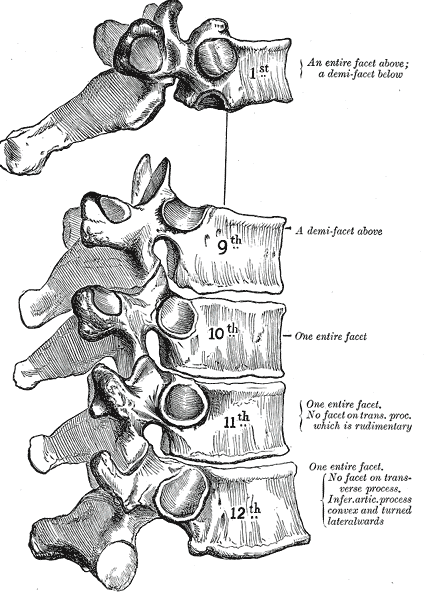
They are generally labeled T1 through T12 in humans with T1 being closer to the neck and T12 more longer than the body. These vertebrae are readily identifiable because they articulate with the ribs and have facets on the sides of their bodies to allow this. The vertebrae are illustrated in the below image.
As with other vertebrae the thoracic vertebrae are so named because of their location. The spinal column formed by the thoracic vertebrae protrudes posteriorly to form the convex thoracic curve of the spine. It doesnt reach the spine until the end of the thorax so what is directly beneath the thoracic vertebrae is the chest cavity.
- The first looks much like C7 and the last four show a progression toward lumbar vertebral structure. General functions of the vertebral column. They are numbered T1-T12.
There are usually 18 of them and they are really interesting vertebrae. They exhibit costal facets on each side at the junction of the vertebral body and the pedicle and on the transverse processes. The spinal curvatures in the thoracic and sacral regions are referred to as primary curvatures because they are present when we are born.
Moving back the next section is the thoracic vertebrae named because they overlie the thorax or chest cavity. Motion palpation assessment of the thoracic spine into extension. - 12 thoracic vertebrae T1-T12 all articulate attached with the ribs - The thoracic vertebrae increase in size from the first to the last.
They increase in size going towards the lumbar vertebrae with the lower ones being a lot larger than the upper. - provides a pivot point for motion and support of the head at the cervical region. - Because it is a multijointed rod the motions of the column occur due to the combined motions of individual vertebrae.
The diaphragm the thin muscle between the thorax and abdomen slants forward-and-down and backward-and-up. AP Unit 5 Set. The anatomy of a typical thoracic vertebra is shown in Figure 74A.
The combing form thoraco means the thorax or chest. It consists of the ribs the sternum and the thoracic vertebrae to which the ribs articulate. The human rib cage is made up of 12 paired rib bones.
Because thoracic rigidity can cause dysfunctional compensations elsewhere if thoracic hypomobility is identified it is important to then assess for all of the possible related conditions. The thoracic vertebrae provide an attachment site for the true ribs and make up part of the back of the chest thorax. The seventh cervical vertebra the prominent vertebra so named because of its long spiny projection adjoins the first thoracic vertebra.
There are twelve pairs of ribs. In vertebrates thoracic vertebrae compose the middle segment of the vertebral column between the cervical vertebrae and the lumbar vertebrae. The thoracic vertebrae provide an attachment site for the true ribs and make up part of the back of the chest thorax.
How many true ribs do humans have. The number is the same in both males and females. So they are called Floating Ribs.
THORACIC CAGE The sternum ribs and thoracic vertebrae make up the bony thorax. Each pair articulates with a different thoracic vertebra. Reproduced with permission from Joseph E.
These vertebrae are readily identifiable because they articate with ribs and have aspects on the sides of their bodies to allow this. It is named for the Atlas of Greek mythology because it supports the globe of the head which is the skull. The thoracic cage also called the rib cage surrounds and protects the heart and lungs in the thoracic cavity.
- maintains the longitudinal axis of the body. Lies at the level of the ninth thoracic vertebra. The atlas and axis are specialized to allow a greater range of motion than normal vertebrae.
Each thoracic vertebra is named for its position within the spine from the first thoracic vertebra T1 on the superior end to the twelfth thoracic vertebra T12 on the inferior end. Each are symmetrically paired on a right and left side. Like typical vertebrae they are separated by intervertebral discs.
Ribs true false floating. HORSES typically have a total of 18 thoracic vertebrae and 18 sets of ribs while CATTLE have a total of 13 thoracic vertebrae and 13 sets of ribs. Artwork by Giovanni Rimasti.
- The weight of the head shoulder girdle upper. The seventh cervical vertebra the prominent vertebra so named because of its long spiny projection adjoins the first thoracic vertebra. The remaining ribs are false ribs so named because their distal attachment is to the costochondral cartilage of their superior neighbor.

Thoracic Vertebrae An Overview Sciencedirect Topics
:watermark(/images/watermark_only_sm.png,0,0,0):watermark(/images/logo_url_sm.png,-10,-10,0):format(jpeg)/images/anatomy_term/thoracic-vertebrae/1fBUPWbpvDDwMRYg92FLRw_j7LbfxmYrP_Vertebrae_thoracicae_02.png)
No comments for "Thoracic Vertebrae Are So Named Because They"
Post a Comment